Author Affiliations
Abstract
1 State Key Laboratory of Precision Electronics Manufacturing, Technology and Equipment, Guangdong University of Technology, Guangzhou 510006, China
2 Key Lab of Optic-Electronic and Communication, Jiangxi Science and Technology, Normal University Nanchang 330038, China
Photoacoustic microscopy (PAM), due to its deep penetration depth and high contrast, is playing an increasingly important role in biomedical imaging. PAM imaging systems equipped with conventional ultrasound transducers have demonstrated excellent imaging performance. However, these opaque ultrasonic transducers bring some constraints to the further development and application of PAM, such as complex optical path, bulky size, and difficult to integrate with other modalities. To overcome these problems, ultrasonic transducers with high optical transparency have appeared. At present, transparent ultrasonic transducers are divided into optical-based and acoustic-based sensors. In this paper, we mainly describe the acoustic-based piezoelectric transparent transducers in detail, of which the research advances in PAM applications are reviewed. In addition, the potential challenges and developments of transparent transducers in PAM are also demonstrated.
Photoacoustic microscopy transparent ultrasound transducer LiNbO3 PMN-PT PVDF CMUT Journal of Innovative Optical Health Sciences
2023, 16(5): 2330001
Author Affiliations
Abstract
1 MOE Key Laboratory of Laser Life Science & Institute of Laser Life Science, College of Biophotonics, South China Normal University, Guangzhou 510631, China
2 Guangdong Provincial Key Laboratory of Laser Life Science, College of Biophotonics, South China Normal University, Guangzhou 510631, China
3 Guangzhou Key Laboratory of Spectral Analysis and Functional Probes, College of Biophotonics, South China Normal University, Guangzhou 510631, China
4 e-mail: xingda@scnu.edu.cn
5 e-mail: qinghuan@scnu.edu.cn
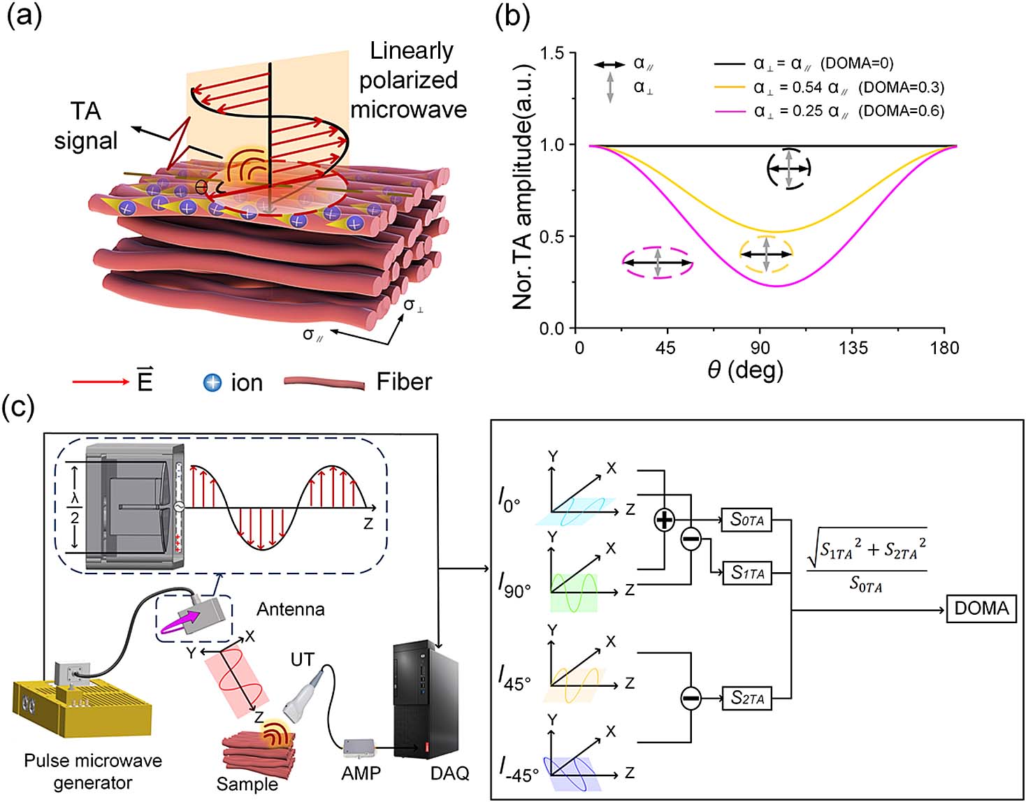
Polarization optical imaging can be used to characterize anisotropy in biological tissue microstructures and has been demonstrated to be a powerful tool for clinical diagnosis. However, the approach is limited by an inability to image targets deeper than due to strong optical scattering in biological tissues. As such, we propose a novel polarization microwave-induced thermoacoustic imaging (P-MTAI) technique to noninvasively detect variations in deep tissue by exploiting the thermoacoustic signals induced by four pulsed microwaves of varying polarization orientations. The proposed P-MTAI method overcomes the penetration limits of conventional polarization optical imaging and provides submillimeter resolution over depths of several centimeters. As part of the paper, the structural characteristics of tissues were quantified using a new parameter, the degree of microwave absorption anisotropy. P-MTAI was also applied to the noninvasive detection of morphological changes in cardiomyocytes as they transitioned from ordered to disordered states, providing a potential indication of myocardial infarction.
Photonics Research
2022, 10(5): 05001297

Author Affiliations
Abstract
1 MOE Key Laboratory of Laser Life Science & Institute of Laser Life Science, South China Normal University, Guangzhou 510631, China
2 College of Biophotonics, South China Normal University, Guangzhou 510631, China
Photoacoustic (PA) microscopy comes with high potential for human skin imaging, since it allows noninvasively high-resolution imaging of the natural hemoglobin at depths of several millimeters. Here, we developed a PA microscopy to achieve high-resolution, high-contrast, and large field of view imaging of skin. A three-dimensional (3D) depth-coding technology was used to encode the depth information in PA images, which is very intuitive for identifying the depth of blood vessels in a two-dimensional image, and the vascular structure can be analyzed at different depths. Imaging results demonstrate that the 3D depth-coded PA microscopy should be translated from the bench to the bedside.
170.5120 Photoacoustic imaging 170.0110 Imaging systems 170.3880 Medical and biological imaging Chinese Optics Letters
2018, 16(8): 081701
华南师范大学, 生物光子学研究院激光生命科学研究院, 暨激光生命科学教育部重点实验室, 广东 广州 510631
报道了一种利用直线电机连续-步进的扫描方式进行光声显微成像的系统, 该系统在运动时走弓字型路线, 其中直线电机在X轴方向上连续运动, 在Y轴方向上以步进的方式运动, 采集卡只在X轴电机运动的过程中连续采集。该成像系统较之前振镜扫描的方式扫描的范围更大, 可达到厘米尺度范围内的生物组织光声成像; 较之前的步进电机逐点扫描的方式扫描速度明显提高。同时本文采用电机带动光和超声换能器一同扫描的方式, 较光和超声换能器不动电机带动样品扫描的方式更灵活。另外利用包埋碳丝的模拟样品和在体小鼠耳朵血管来验证系统的成像能力。实验结果表明, 这种快速光声显微成像方法及其系统可以实现在体组织的高分辨率成像, 有望成为一种无创、实时的光声显微镜应用于生物医学当中。
连续-步进运动 快速光声成像 光声显微镜 continuous-stepping motion fast photoacoustic imaging photoacoustic microscope


